
The Modality Matrix Series Portfolio
This sixth installment explores the specialized domain of Targeted Radionuclide Therapies (TRTs) and theranostics. Transitioning these dual-modality assets into first-in-human clinical trials requires an advanced quantitative Clinical Pharmacology Plan (CPP) capable of integrating radiation dosimetry, radioactive decay kinetics, and multi-isotope PBPK modeling.
| Article Sequence | Thematic Focus | Status |
|---|---|---|
| Article 1 | Overview of Modalities and the Clinical Pharmacology Plan | Published |
| Article 2 | Small Molecule PK and Bioavailability | Published |
| Article 3 | Large Molecule Biologics and Subcutaneous Delivery | Published |
| Article 4 | Antibody-Drug Conjugates (ADCs) and Multi-Analyte PK | Published |
| Article 5 | Oligonucleotide Therapeutics (ASOs, siRNAs, LNPs) | Published |
| Article 6 | Targeted Radionuclide Therapies (TRTs) and Theranostics | Current Report |
Introduction: The Theranostic Renaissance & The Geometric vs. Pharmacologic Divide
The clinical landscape of precision oncology is experiencing a fundamental shift driven by Targeted Radionuclide Therapies (TRTs) and the theranostic paradigm—the systematic pairing of a diagnostic imaging surrogate with a therapeutic cytotoxic radionuclide using the same molecular targeting vector. By enabling drug developers to visually confirm target expression, quantify whole-body biodistribution, and calculate localized tissue radiation deposition prior to therapeutic dosing, theranostics represents the ultimate realization of personalized medicine.
However, transitioning a novel radiopharmaceutical from preclinical proof-of-concept to a first-in-human (FIH) Phase 1 clinical trial presents a profound clinical pharmacology challenge. To succeed, biotech sponsors must first unlearn the development frameworks associated with both traditional chemotherapies and traditional radiotherapies [1].
The Top-Down Geometric vs. Bottom-Up Pharmacologic Paradigm
Historically, radiation oncology has been dominated by External Beam Radiation Therapy (EBRT) and localized brachytherapy. The clinical development and dosing of traditional EBRT is fundamentally a top-down, geometric physics exercise. Radiation is delivered from an external linear accelerator to a statically mapped, macroscopically defined anatomical coordinate space via CT/MRI treatment planning. Dosing is prescribed as fixed physical energy increments (e.g., 2 Gy fractions to a total biological target of 60 Gy). Because the radiation source is external and non-chemical, systemic pharmacokinetics, target receptor density, cellular internalization rates, and physiological clearance organs play zero role in dose delivery.
In stark contrast, a Targeted Radionuclide Therapy is a systemically administered chemical entity. Its development is a bottom-up, dynamic pharmacological exercise. Once infused into systemic circulation, the radiation source is mobile. Radiation delivery is governed entirely by molecular biodistribution, vascular extravasation, Target-Mediated Drug Disposition (TMDD), intracellular endocytosis, and physiological excretion pathways such as megalin/cubilin reabsorption in renal proximal tubules [2].
Furthermore, drug developers cannot simply extrapolate traditional EBRT normal-organ dose limits (such as a 23 Gy renal limit) directly to TRTs. EBRT delivers high-intensity, instantaneous physical dose rates over minutes. TRTs deliver low-dose-rate, continuous, exponentially decaying radiation over days or weeks, exhibiting completely different cell-repair kinetics and relative biological effectiveness (RBE). Therefore, while an EBRT program requires a medical physicist, a TRT asset requires an expert Quantitative Clinical Pharmacologist and DMPK strategist capable of modeling radioactive decay kinetics, effective tissue half-lives (t1/2,eff), Linear Energy Transfer (LET), and cellular-level absorbed radiation doses.
The Regulatory Shift: The Fall of Empirical Dosing
This pharmacological reality has triggered a major regulatory tightening. Under the FDA Oncology Center of Excellence (OCE) Project Optimus initiative and recent dosage optimization guidances specifically tailored to radiopharmaceuticals, the era of deploying empirical, legacy 3+3 dose-escalation designs that administer fixed activities (e.g., 6.8 GBq every 6 weeks) without mechanistic dosimetric justification is closing [3]. For emerging biotech sponsors, securing an Investigational New Drug (IND) approval and avoiding clinical holds now requires a Quantitative Clinical Pharmacology Plan (CPP) that integrates molecular imaging, dynamic blood-time curve modeling, and organ-level radiation dosimetry from day one.
Consultant’s Strategic Advisory Note — The “EBRT Extrapolation” Trap: In early advisory meetings, we frequently see startup executive teams assume that if a radioligand delivers an absorbed renal dose below the classic EBRT threshold of 23 Gy, renal safety is guaranteed. Do not make this assumption before regulatory agencies. Because TRTs concentrate intracellularly within proximal tubule cells and deliver continuous, un-fractionated radiation over hundreds of hours, nephrotoxicity can occur at absorbed doses that look deceivingly safe on an EBRT chart—especially with Targeted Alpha Therapies (TATs). Your Phase 1 Clinical Pharmacology Plan must treat radiation dosimetry as a dynamic pharmacological parameter, establishing custom, model-informed exposure-safety thresholds specific to your vector’s clearance kinetics.
Section 1: Anatomy of a Radiopharmaceutical Conjugate Across Targeting Vectors
To engineer a robust clinical pharmacology strategy, drug developers must deconstruct the radiopharmaceutical into its four core functional domains: the Targeting Vector, the Pharmacokinetic Linker, the Chelator, and the Radionuclide.
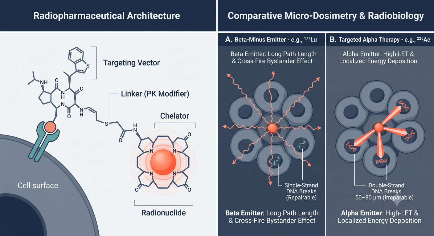
Figure 1: Modular Radiopharmaceutical Architecture & Comparative Beta vs. Alpha Micro-Dosimetry
Comparative Vector Pharmacology: Peptides & Small Molecules vs. Macromolecules
The choice of targeting vector dictates the macroscopic biodistribution, systemic clearance rate, and dose-limiting toxicity (DLT) profile of the asset:
- Small Molecule & Peptide Conjugates (The Startup Standard): Constructing TRTs using small molecules (e.g., PSMA inhibitors) or cyclic peptides (e.g., somatostatin receptor analogs like OCTREOTATE) dominates the current biotech pipeline. Due to their low molecular weight (<1.5 kDa to 5 kDa), these vectors display rapid vascular extravasation and exceptional solid tumor penetration. Systemically, they undergo rapid renal clearance, causing plasma radioactivity levels to plummet within hours of intravenous infusion [4]. While this protects red bone marrow from prolonged circulating radiation exposure, it shifts the primary toxicological burden directly to the kidneys and specialized epithelial tissues like salivary glands.
- Macromolecules / Intact mAbs (Radioimmunotherapy – RIT): Utilizing intact monoclonal antibodies (∼150 kDa) represents a legacy approach with limited commercial success in solid tumors. Because intact immunoglobulins exceed the glomerular filtration threshold and undergo FcRn-mediated recycling, they display prolonged serum half-lives (t1/2 > 1 to 3 weeks). While this prolongs tumor exposure, it forces circulating blood and red bone marrow to absorb massive, continuous radiation doses. Consequently, myelosuppression becomes the primary dose-limiting toxicity, often occurring long before therapeutic radiation doses can be delivered to solid tumors.
| Targeting Vector Modality | Systemic Half-Life & Extravasation | Primary Clearance & DLT Sink | Strategic Consultant Take |
|---|---|---|---|
| Small Molecule Ligands (e.g., PSMA-617) |
Extremely short plasma t1/2 (<24 hr). Rapid, uniform solid tumor diffusion. | Renal filtration. DLTs: Salivary/lacrimal glands (xerostomia) and renal cortex. | Industry standard for solid tumors. Demands proactive mitigation for off-target salivary uptake. |
| Cyclic Peptides (e.g., DOTATATE) |
Short plasma t1/2 (∼48–72 hr terminal). Rapid tissue penetration. | Megalin/cubilin renal reabsorption. DLT: Renal cortex radiation nephritis. | Highly efficient tumor delivery. Requires basic amino acid co-infusion for kidney protection. |
| Intact Antibodies (RIT) (e.g., Legacy mAbs) |
Prolonged plasma t1/2 (1–3 weeks). Slow vascular extravasation. | Hepatic/RES catabolism. DLT: Red bone marrow myelosuppression. | High circulating toxicity burden. Best reserved for hematologic cancers or pre-targeted platforms. |
Table 1: Comparative DMPK & Toxicity Profiles Across Targeting Vectors
Isotope Physics & Radiobiology: Beta Emitters vs. Targeted Alpha Therapies
The therapeutic index of a TRT is fundamentally governed by the nuclear decay physics of the attached radionuclide:
Beta-Minus (β–) Emitters: Isotopic payloads such as Lutetium-177 (177Lu) and Iodine-131 (131I) emit electrons with relatively low Linear Energy Transfer (LET ≈ 0.2–2 keV/μm) and long tissue path lengths (1–10 mm, spanning hundreds of cell diameters). Beta emitters cause cellular apoptosis primarily through indirect free-radical generation and single-strand DNA breaks, which hypoxic or radiation-resistant tumors can often repair. However, their long path length creates a potent bystander effect, making them highly effective for treating large, heterogeneous tumor masses where antigen expression is patchy [5].
Targeted Alpha Therapies (TATs): Alpha emitters—such as Actinium-225 (225Ac), Lead-212 (212Pb), and Astatine-211 (211At)—emit massive helium nuclei (2 protons, 2 neutrons) characterized by exceptionally high Linear Energy Transfer (LET ≈ 80–100 keV/μm) and a very short tissue path length (50–80 μm, spanning roughly 2–10 cell diameters). As an alpha particle traverses a cell, it deposits immense localized kinetic energy, generating dense, irreparable double-strand DNA breaks. This cytotoxic mechanism is entirely independent of tissue oxygenation or cell-cycle stage, overcoming classic beta-resistance and allowing for the precision eradication of micro-metastases without irradiating adjacent healthy tissue.
The Theranostic “Matched Pair” DMPK Nuance
A critical point of failure in early clinical pharmacology planning is assuming that a diagnostic imaging conjugate and a therapeutic conjugate share identical biodistribution and elimination kinetics. In clinical practice, developers frequently rely on diagnostic surrogates, such as pairing Diagnostic Gallium-68 (68Ga) or Copper-64 (64Cu) for PET imaging with Therapeutic Lutetium-177 (177Lu) or Actinium-225 (225Ac) using the same universal chelator.
While the targeting peptide sequence remains unchanged, substituting a metal ion with a different ionic radius, valence state, and coordination number alters the overall chelate geometry, surface charge density, and lipophilicity of the molecule. This structural perturbation frequently creates a diagnostic-to-therapeutic PK disconnect. A 68Ga-labeled peptide may display a lower equilibrium dissociation constant (Kd), different plasma protein binding, or higher hepatic clearance than its 177Lu-labeled counterpart. Using imaging data from a chemically divergent surrogate to calculate predictive organ dosimetry for the therapeutic isotope can introduce error rates exceeding 20–30% [4].
To overcome this liability, the vanguard of the radiopharmaceutical industry is shifting toward True Elemental Matched Pairs. These pairs utilize different radiocobalt, radioiodine, or radiolead isotopes of the exact same chemical element (such as Lead-203 for SPECT imaging paired with Lead-212 for Targeted Alpha Therapy, or Copper-64 paired with Copper-67). Because the chemical element is identical, the coordination chemistry, lipophilicity, receptor binding affinity, and in vivo biodistribution are mathematically and biologically indistinguishable, providing absolute DMPK fidelity between the diagnostic imaging cohort and the therapeutic dosing cohort.
| Theranostic Pair Type | Representative Isotope Pairs | Coordination Chemistry & Kd | Phase 1 Dosimetry Accuracy |
|---|---|---|---|
| Diagnostic Chemical Surrogates | 68Ga (PET) or 64Cu (PET) paired with 177Lu (Beta) or 225Ac (Alpha). | Divergent: Different ionic radii alter chelator cage geometry and shift target affinity by 2- to 10-fold. | Error-Prone: Extrapolating therapeutic organ doses from a chemical surrogate can introduce 20–30% dosimetric error. |
| True Elemental Matched Pairs | 203Pb (SPECT) / 212Pb (Alpha) 64Cu (PET) / 67Cu (Beta) 123I (SPECT) / 131I (Beta) |
Identical: Same chemical element guarantees identical oxidation states, chelator geometry, and Kd. | Gold Standard: Provides 1:1 predictive accuracy, dramatically de-risking FIH dosage optimization under Project Optimus. |
Table 2: Theranostic Matched Pairs — Diagnostic Surrogates vs. True Elemental Pairs
Section 2: Approved Clinical Benchmarks & Lessons Learned
Analyzing the clinical pharmacology data of the industry’s blockbuster approvals reveals both the therapeutic potential and the pharmacokinetic limitations of first-generation TRTs:
- Lutathera (177Lu-DOTATATE): Approved for somatostatin receptor-positive gastroenteropancreatic neuroendocrine tumors (GEP-NETs), Lutathera utilizes an octreotide peptide derivative. Its DMPK profile is defined by rapid biexponential blood clearance (t1/2,α ≈ 1.6 hours; terminal t1/2,β ≈ 71 hours) and primary urinary excretion (>60% cleared within 24 hours). Because the peptide is freely filtered by the glomerulus and reabsorbed by megalin/cubilin endocytic receptors in the proximal tubule, the kidneys represent the primary dose-limiting organ [5].
- Pluvicto (177Lu-PSMA-617): Approved for metastatic castration-resistant prostate cancer (mCRPC), Pluvicto pairs a small molecule glutamate-urea-lysine PSMA inhibitor with Lutetium-177. It exhibits an even more rapid initial vascular clearance than peptide systems, distributing rapidly into PSMA-expressing tumor tissues and specialized non-target glands. Salivary and lacrimal gland uptake drives xerostomia (dry mouth), which, alongside cumulative bone marrow toxicity from circulating retention, dictates clinical tolerability [5].
The Problem with Legacy Empirical Dosing
Both Lutathera and Pluvicto were approved on fixed, empirical activity schedules (4 doses of 7.4 GBq for Lutathera; 6 doses of 6.8 GBq for Pluvicto). While administratively convenient for multicenter Phase 3 trials, this one-size-fits-all paradigm represents suboptimal clinical pharmacology. In real-world populations, inter-patient variability in renal clearance, tumor volume, and receptor density causes absorbed radiation doses to span a 5-fold to 10-fold range across patients receiving the exact same administered MBq activity [3].
Fixed empirical dosing under-doses patients with high tumor sinks or rapid clearance (leaving survival benefits on the table) while over-dosing patients with mild renal impairment or low tumor volumes, driving premature treatment discontinuation due to myelosuppression or nephrotoxicity. Next-generation biotechs must compete by demonstrating that individualized, dosimetry-guided dosing expands the therapeutic window beyond what Pluvicto and Lutathera achieved.
Consultant’s Strategic Advisory Note — The Cold-Mass / Specific Activity Dilemma: In early Phase 1 dose escalation, biotechs frequently miscalculate the ratio of radiolabeled drug to unlabeled drug—a parameter known as Specific Activity (MBq/μg or GBq/μmol). When formulating a drug, a significant mass of unlabeled (“cold”) targeting peptide or small molecule is inevitably present alongside the hot conjugate. If your total peptide mass (cold + hot) exceeds the saturation threshold of the target receptor pool in the tumor, the cold molecules will outcompete the radiolabeled molecules for tumor binding sites. This creates a catastrophic Phase 1 artifact: as you escalate the radiation dose by adding more chemical mass, tumor uptake drops, systemic clearance slows, and healthy organ toxicity spikes. Your CPP must mandate explicit in vivo receptor saturation modeling to define the maximum allowable chemical mass per cohort before dosing your first human subject.
Section 3: The TRT PK/PD Disconnect & Dosimetry-Guided Dosing
To execute a modern radiopharmaceutical development program, DMPK scientists must reconcile the profound disconnect between systemic blood kinetics and tissue-level pharmacodynamics. In conventional drug development, plasma drug concentration over time serves as a direct surrogate for receptor occupancy and clinical efficacy. In radionuclide therapy, systemic blood clearance is merely the transport phase.
Once a radiotheranostic enters a tumor cell or normal organ cell via receptor-mediated internalization, the chemical vehicle is often degraded, but the radioactive isotope remains trapped intracellularly, continuing to decay according to its physical half-life (t1/2,phys). Consequently, plasma elimination half-life does not correlate with organ-absorbed dose or antitumor efficacy. A peptide may clear systemic circulation within 120 minutes, yet generate an effective tissue half-life (t1/2,eff) of 100+ hours within the renal cortex or tumor matrix.
The Mechanics of Internal Dosimetry: From MBq to Grays (Gy)
To evaluate efficacy and toxicity quantitatively, the clinical pharmacology team must convert administered activity (MBq or GBq) into tissue-absorbed radiation dose, expressed in Grays (Gy), where 1 Gy = 1 Joule of radiation energy deposited per kilogram of tissue. This requires a formal dosimetry workflow:
- Image Acquisition: Following the administration of a diagnostic tracer or a trace-labeled therapeutic dose, serial quantitative imaging (PET/CT or SPECT/CT) is performed across multiple time points (e.g., 1, 4, 24, 72, and 168 hours post-injection).
- Time-Activity Curve (TAC) Generation: Volumes of Interest (VOIs) are segmented over tumors and critical clearance organs (kidneys, liver, red marrow, salivary glands). The activity within each organ is plotted over time to construct tissue-specific TACs.
- Time-Integrated Activity (Ã): Mathematical integration of the TAC from time zero to infinity yields the Time-Integrated Activity (formerly termed cumulated activity), which represents the total number of nuclear disintegrations that occurred within that source organ:
à = ∫ A(t) dt - MIRD & Voxel-Based Dose Calculation: Using the formalisms established by the Medical Internal Radiation Dose (MIRD) committee, the absorbed dose (D) to a target tissue from radiation emitted by a source organ is calculated by multiplying the Time-Integrated Activity by a radionuclide- and geometry-specific factor known as the S-value:
D(target) = ∑ [ Ã(source) × S(target ← source) ]
While standard MIRD formalisms rely on generalized anthropomorphic phantom models, advanced Phase 1 programs now deploy Voxel-Based Dosimetry. This technique maps S-values directly onto patient-specific CT densities at the 3D voxel level, capturing localized radiation hot-spots within heterogeneous tumor masses or the renal cortex. By anchoring the Phase 1 Clinical Pharmacology Plan to voxel-based dosimetric endpoints (Gy/GBq), sponsors can mathematically prove to regulatory authorities that a proposed Phase 2 dose delivers a tumor-absorbed dose exceeding the oncogenic threshold (>50–60 Gy) while constraining renal cortex exposure below biological tolerance limits (<23 Gy for beta emitters).
Section 4: The Quantitative Engine: Integrating Blood PK with Imaging Dosimetry
To build a Quantitative Systems Pharmacology (QSP) or Model-Informed Drug Development (MIDD) platform for a Targeted Radionuclide Therapy, DMPK scientists must synthesize two distinct data streams: serial blood/plasma radioactivity measurements and longitudinal volumetric quantitative imaging. While traditional pharmacometrics relies almost exclusively on plasma sampling to infer peripheral compartment distribution, TRT development allows us to directly observe and quantify tissue-level drug disposition in real time [3].
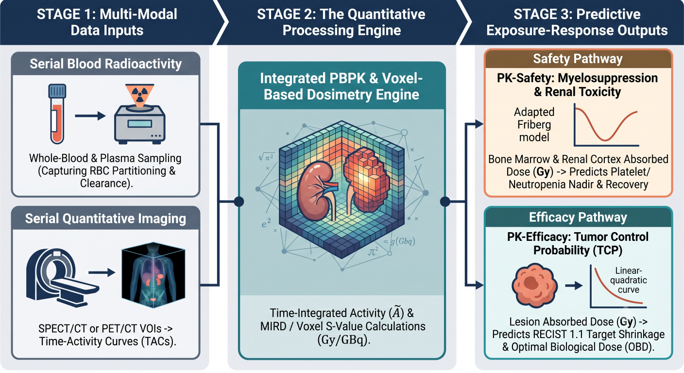
Figure 2: The TRT Quantitative Modeling Engine — From Blood & Imaging to Predictive PK-Safety and PK-Efficacy
1. PK-Safety Modeling: Adapting the Friberg Myelosuppression Model
Hematological toxicity—specifically Grade 3/4 thrombocytopenia, neutropenia, and leukopenia—is a primary dose-limiting toxicity across both beta and alpha radiopharmaceuticals. To predict and mitigate myelosuppression, clinical pharmacologists must adapt the classical Friberg semi-mechanistic hematological toxicity model.
In standard chemotherapy modeling, the Friberg framework links plasma drug concentration to the inhibition of proliferative bone marrow stem cells. In radiopharmaceutical pharmacometrics, circulating chemical mass concentration is biologically irrelevant; myelosuppression is driven by red marrow absorbed radiation dose rate (D̊marrow) or cumulative Time-Integrated Activity. By replacing chemical concentration with bone marrow absorbed dose rate, the system of differential equations governing circulating blood counts becomes:
d(Prolim)/dt = ktr × Prolim × [ 1 – Slope × D̊marrow(t) ] × (Circ0 / Circ)γ – ktr × Prolim
d(Circ)/dt = ktr × Transitn – ktr × Circ
Where Prolim represents the proliferative stem cell compartment, Circ represents circulating mature blood cells, ktr is the inter-compartmental transit rate constant, and γ is the feedback exponent accounting for endogenous rebound. Integrating this adapted Friberg model allows sponsors to predict the nadir timing, severity, and recovery trajectory of leukocytes and platelets across proposed Phase 1 activity escalation cohorts, proving to regulatory agencies that a proposed dose increment will not trigger irreversible myeloablation.
2. PK-Efficacy Modeling: Tumor Control Probability (TCP)
To justify dosage optimization under FDA Project Optimus, sponsors must establish quantitative PK-Efficacy models that correlate tumor-absorbed radiation dose with longitudinal target lesion shrinkage (RECIST 1.1 criteria) and Overall Survival. Instead of using plasma AUC, pharmacometricians utilize Tumor Control Probability (TCP) formalisms and linear-quadratic (LQ) radiobiological models. The surviving fraction (SF) of tumor cells within a segmented lesion following a cumulative absorbed dose (Dtumor) delivered at an exponentially decaying dose rate (λ) is modeled as:
SF = exp( -α × Dtumor – β × G(λ) × Dtumor2 )
Where α and β are tissue-specific radiosensitivity coefficients (representing single-hit kill vs. dual-hit repairable damage), and G(λ) is the Lea-Catcheside dose-protraction factor accounting for DNA repair during the prolonged low-dose-rate exposure typical of TRTs. By mapping these survival fractions against longitudinal tumor growth inhibition models—benchmarked against post-hoc analyses of landmark trials like VISION (Pluvicto) and NETTER-1 (Lutathera)—sponsors can mathematically identify the Optimal Biological Dose (OBD) prior to initiating large-scale Phase 2 trials.
Consultant’s Strategic Advisory Note — The “Whole-Blood vs. Plasma” Gamma Counting Error: During early Phase 1 execution, clinical sites frequently collect plasma samples for gamma counting while discarding the cellular blood pellet. Do not allow your bioanalytical team to make this omission. Radiolabeled peptides and small molecules often display significant partitioning into red blood cells (RBCs) or non-specific binding to cellular membranes. If you calculate blood-based red marrow dosimetry using plasma radioactivity alone, you will systematically underestimate circulating blood retention and bone marrow absorbed dose, leading to unexpected Phase 1 cytopenias that could trigger a clinical hold. Your Clinical Pharmacology Plan must mandate both whole-blood and plasma scintillation/gamma counting at every pharmacokinetic sampling time point.
Section 5: Modality-Specific DMPK & Toxicity Mitigation Strategies
Because radiopharmaceuticals are systemic chemical vectors carrying nuclear payloads, their toxicological profile is governed by anatomical clearance sinks and radioactive decay chains. A competitive CPP must deploy proactive pharmacological strategies to mitigate these modality-specific liabilities.
1. The Renal Sink & Competitive Amino Acid Co-Infusion
For small molecule and peptide TRTs, the kidneys represent the primary clearance organ and the predominant dose-limiting toxicity sink. As low-molecular-weight vectors pass through the glomerulus, they are reabsorbed from the glomerular filtrate by specialized endocytic receptors—primarily megalin and cubilin—located on the apical membrane of the proximal tubule epithelial cells. Once endocytosed, the vector is degraded in lysosomes, but the radiometal is retained within the tubular epithelium, irradiating the renal cortex over its entire physical half-life [2].
To mitigate nephrotoxicity and expand the therapeutic window, clinical pharmacology protocols must incorporate competitive renal protection. Infusing high-dose basic amino acids—specifically L-lysine and L-arginine—prior to and during radiopharmaceutical administration saturates the megalin/cubilin receptor network. This competitive blockade inhibits tubular reabsorption by 30–50%, flushing the unbound radiopharmaceutical directly into the urine and lowering renal cortex absorbed dose without impacting high-affinity tumor receptor binding.
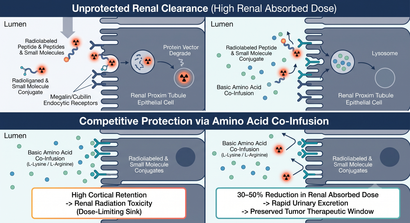
Figure 3: Megalin/Cubilin Renal Reabsorption & Competitive Blockade via Basic Amino Acid Co-Infusion
2. Salivary & Lacrimal Gland Toxicity: The PSMA Challenge
Xerostomia (severe dry mouth) and xerophthalmia (dry eyes) represent the primary quality-of-life dose-limiting toxicities for PSMA-targeted therapies. Unlike tumor uptake, which is driven by high-affinity catalytic site binding on prostate-specific membrane antigen, salivary gland accumulation is complex and multifactorial, involving both non-specific pinocytosis and low-level physiological PSMA expression in salivary ductal cells [5].
Because basic amino acid infusions do not block salivary uptake, sponsors must explore localized DMPK mitigation strategies. These include sialendoscopy, localized botulinum toxin injections to induce temporary glandular apoptosis prior to dosing, or topical cooling (ice packs) applied to the parotid and submandibular glands during infusion to vasoconstrict local blood flow and reduce vascular drug delivery to the glands.
3. The “Daughter Isotope Recoil” Problem in Targeted Alpha Therapies
As biotechs pivot from beta emitters (177Lu) to Targeted Alpha Therapies (225Ac, 212Pb, 211At), clinical pharmacologists face a unique radiochemical phenomenon: Daughter Isotope Recoil. Alpha decay is not a single-step process; isotopes like Actinium-225 undergo a nuclear decay chain generating multiple alpha- and beta-emitting daughter radioisotopes before reaching a stable state:
225Ac → [221Fr] → [217At] → [213Bi] → [209Pb] → 209Bi (Stable)
When an Actinium-225 nucleus ejects an alpha particle at ∼6 MeV, conservation of momentum dictates that the remaining daughter nucleus (221Fr) experiences a powerful kinetic recoil energy of approximately 100 keV. To put this energy into chemical perspective: the covalent and thermodynamic coordination bonds securing a radiometal within an advanced chelator complex (such as DOTA or MACROPA) possess dissociation energies of only 3–5 eV. The recoil kinetic energy exceeds the chelator bond strength by a factor of 20,000 [6].
Consequently, every alpha decay instantaneously shatters the chemical chelate bond, physically ejecting the radioactive daughter isotope out of the targeting vector. Once liberated from the vector, these unchelated daughter isotopes (such as Francium-221 and Bismuth-213) behave as free chemical elements. They are swept away from the tumor site by blood flow and re-distribute according to their own elemental pharmacology—with Bismuth-213 accumulating heavily in the renal cortex and Francium/Lead isotopes tracking to bone marrow. A competitive TAT Clinical Pharmacology Plan must incorporate multi-isotope PBPK modeling to mathematically prove that the cumulative radiation burden from detached daughter isotopes will not cause delayed nephrotoxicity or irreversible marrow failure.
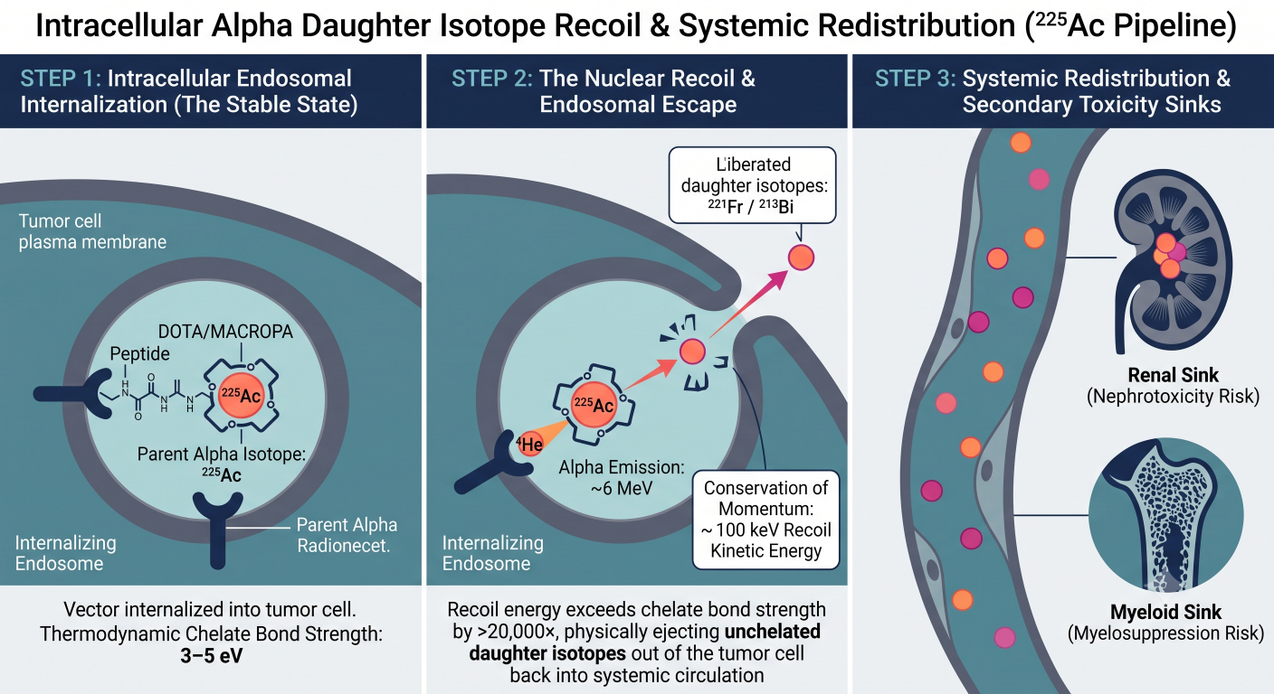
Figure 4: Intracellular Alpha Daughter Isotope Recoil & Systemic Redistribution in Actinium-225 Pipelines
Section 6: Designing the TRT Clinical Pharmacology Plan (CPP)
A comprehensive TRT Clinical Pharmacology Plan must harmonize traditional regulatory pharmacology requirements with the specialized physical realities of nuclear medicine.
1. DDI Strategy: Moving Beyond CYP450 to Receptor Competition
Traditional small molecule and biologic drug-drug interaction (DDI) programs focus heavily on Cytochrome P450 (CYP3A4, CYP2D6) metabolism and transporter inhibition (P-gp, OATP1B1). For radiopharmaceuticals, traditional CYP/transporter DDI liabilities are practically negligible. Because TRTs are administered at exceptionally low chemical mass doses—often in the low microgram range, generating transient circulating plasma concentrations in the low picomolar or nanomolar range—they do not achieve the molar concentrations required to competitively inhibit or induce hepatic CYP enzymes [1].
However, the CPP must aggressively evaluate Receptor-Competition and Axis-Modulating DDIs:
- Cold-Mass Competition: Co-administering medications that share the same transport or receptor pathway (e.g., unlabeled somatostatin analogs like octreotide LAR administered too close to Lutathera dosing) will competitively block tumor receptor binding, drastically reducing therapeutic radiation delivery.
- Target Upregulation / Modulation: In PSMA theranostics, co-administering Androgen Receptor Pathway Inhibitors (ARPIs; e.g., enzalutamide, abiraterone) temporarily upregulates PSMA gene transcription and surface expression on prostate cancer cells. A strategic CPP can exploit this pharmacology, using transient ARPI co-dosing to deliberately boost tumor radiation uptake prior to radiotherapy administration.
- Gastrointestinal / pH Modulators: For orally administered or gastrointestinally reabsorbed vectors, proton pump inhibitors (PPIs) or agents altering intraluminal pH can significantly modify systemic absorption and clearance kinetics.
2. Organ Impairment Trials in Nuclear Medicine
Conducting dedicated Phase 1 renal or hepatic impairment studies for radiopharmaceuticals presents unique ethical and dosimetric challenges. Because small molecule and peptide TRTs rely heavily on renal filtration, patients with baseline moderate-to-severe renal impairment (eGFR < 45 mL/min/1.73m2) will experience delayed vascular clearance, elevating whole-body background radiation and sharply increasing bone marrow and renal cortex absorbed dose.
Rather than conducting standalone clinical impairment trials—which risk severe radiation toxicity in organ-compromised patients—the FDA and EMA increasingly accept Model-Informed Organ Impairment Strategies. Sponsors deploy population pharmacokinetic (PopPK) and PBPK models parameterized with baseline renal function metrics (CrCl, eGFR, cystatin-C) derived from the broad Phase 1/2 patient population. By simulating the radiation dosimetry of compromised cohorts in silico, sponsors can establish precise, model-derived activity reduction guidelines within the prescribing label without exposing vulnerable patients to empirical dosing experiments [1].
3. Safety Pharmacology: ECG & QTc Monitoring Protocols
The FDA requires an evaluation of cardiac safety and QTc prolongation risk for all novel molecular entities, including radiopharmaceuticals. However, executing standard high-time-resolution ECG monitoring (e.g., triplicate ECGs matched to 10 rapid blood draw time points over 24 hours) faces logistical barriers in a nuclear medicine suite. Drawing frequent radioactive blood samples and attaching telemetry equipment to patients emitting gamma or bremsstrahlung radiation raises radiation exposure risks for clinical nursing staff.
To solve this, the CPP should propose a Concentrations-QTc (C-QTc) Risk-Based Mitigation Strategy. Sponsors first establish in vitro hERG channel assay data demonstrating an exceptionally wide safety margin (>10,000-fold between the therapeutic picomolar Cmax and the hERG IC50 threshold). This is paired with a streamlined clinical ECG protocol utilizing continuous Holter monitoring and sparse, optimized blood sampling (e.g., baseline, end of infusion, and 2 hours post-infusion) during early FIH escalation cohorts, minimizing nurse exposure while mathematically ruling out clinical cardiac liability [1].
Actionable Consulting Deliverable: Phase 1 Clinical Pharmacology & Dosimetry Readiness Checklist
For biotech CSOs and clinical development leaders preparing an IND submission for a novel radiopharmaceutical, this operational checklist serves as an internal Standard Operating Procedure (SOP) reference to verify that all quantitative pharmacology, dosimetry, and regulatory requirements are locked down prior to FIH execution.
| Phase / Domain | Strategic Requirement | Operational Verification Standard | Sign-Off |
|---|---|---|---|
| 1. Vector & Chemistry | Elemental Matched Pair Validation | If using a chemical surrogate (68Ga, 64Cu), binding (Kd) and animal biodistribution must prove DMPK parity with the therapeutic isotope. Priority given to true elemental pairs (203Pb/212Pb or 64Cu/67Cu). | □ Verified |
| 1. Vector & Chemistry | Specific Activity Ceiling | Establish maximum allowable unlabeled (“cold”) ligand mass (μg/dose) via receptor saturation modeling to prevent tumor receptor blockade during FIH escalation. | □ Verified |
| 2. Bioanalysis & PK | Whole-Blood vs. Plasma Counting | Bioanalytical protocol explicitly mandates dual gamma scintillation counting of both whole blood and separated plasma to quantify erythrocyte partitioning and protect marrow dosimetry. | □ Verified |
| 2. Bioanalysis & PK | Metabolite Profiling & Radio-HPLC | Validated Radio-HPLC or Radio-TLC assays established to quantify circulating intact radiolabeled conjugate versus free, unchelated radiometal in plasma and urine. | □ Verified |
| 3. Clinical Dosimetry | Multi-Time-Point Imaging Protocol | Clinical protocol mandates serial quantitative SPECT/CT or PET/CT imaging across a minimum of 4 to 5 time points (e.g., 1–2 hr, 4–6 hr, 24 hr, 72 hr, and 168 hr post-injection). | □ Verified |
| 3. Clinical Dosimetry | Voxel-Based Dosimetry Engine | Software infrastructure validated to compute tissue-absorbed dose (Gy/GBq) at the 3D voxel level for tumors, renal cortex, red marrow, and salivary glands. | □ Verified |
| 4. Pharmacometrics | Project Optimus E-R Justification | CPP includes pre-specified formalisms linking tumor-absorbed dose with RECIST 1.1 shrinkage (TCP models) and red marrow dose with hematological toxicity (adapted Friberg model). | □ Verified |
| 4. Pharmacometrics | Alpha Daughter Recoil PBPK Model | For Targeted Alpha Therapies (TATs) only: PBPK simulation platform parameterized to model the systemic release, redistribution, and secondary organ dosimetry of unchelated daughter isotopes. | □ Verified |
| 5. Trial Design | Renal Protection Co-Infusion SOP | Clinical protocol includes standardized, mandated co-infusion of basic amino acids (e.g., 2.5–5% L-lysine/L-arginine solution initiated 30 minutes prior to TRT infusion). | □ Verified |
| 5. Trial Design | Receptor-Competition DDI Washout | Protocol inclusion/exclusion criteria enforce strict washout windows for competing cold somatostatin analogs (e.g., octreotide LAR washout >4 weeks) or define ARPI co-dosing schedules. | □ Verified |
| 6. Regulatory & Safety | Streamlined C-QTc Cardiac Protocol | High-sensitivity in vitro hERG patch-clamp data submitted alongside a sparse clinical Holter monitoring protocol to minimize clinical staff radiation exposure while satisfying cardiac guidelines. | □ Verified |
Table 3: Phase 1 Clinical Pharmacology & Dosimetry Readiness Checklist
Abbreviations
ARPI: Androgen Receptor Pathway Inhibitor
AUC: Area Under the Curve
CPP: Clinical Pharmacology Plan
CrCl: Creatinine Clearance
CYP: Cytochrome P450
DDI: Drug-Drug Interaction
DLT: Dose-Limiting Toxicity
DMPK: Drug Metabolism and Pharmacokinetics
EBRT: External Beam Radiation Therapy
ECG: Electrocardiogram
eGFR: Estimated Glomerular Filtration Rate
E-R: Exposure-Response
FIH: First-In-Human
GEP-NET: Gastroenteropancreatic Neuroendocrine Tumor
hERG: Human Ether-à-go-go-Related Gene
HPLC: High-Performance Liquid Chromatography
IND: Investigational New Drug
LET: Linear Energy Transfer
mAb: Monoclonal Antibody
mCRPC: Metastatic Castration-Resistant Prostate Cancer
MIDD: Model-Informed Drug Development
MIRD: Medical Internal Radiation Dose
OBD: Optimal Biological Dose
OCE: Oncology Center of Excellence
PBPK: Physiologically Based Pharmacokinetic
PET: Positron Emission Tomography
PopPK: Population Pharmacokinetics
PPI: Proton Pump Inhibitor
PSMA: Prostate-Specific Membrane Antigen
QSP: Quantitative Systems Pharmacology
RBE: Relative Biological Effectiveness
RECIST: Response Evaluation Criteria in Solid Tumors
RES: Reticuloendothelial System
RIT: Radioimmunotherapy
SPECT: Single-Photon Emission Computed Tomography
SSTR: Somatostatin Receptor
TAC: Time-Activity Curve
TAT: Targeted Alpha Therapy
TCP: Tumor Control Probability
TMDD: Target-Mediated Drug Disposition
TRT: Targeted Radionuclide Therapy
VOI: Volume of Interest
Technical References
- U.S. Food and Drug Administration (FDA). (2023). Clinical Pharmacology Considerations for the Development of Radiopharmaceuticals. Guidance for Industry / Oncology Center of Excellence (OCE).
- Vegt, E., de Jong, M., Wetzels, J. F., & Masereeuw, R. (2010). Renal toxicity of radiolabeled peptides and antibody fragments: mechanisms and protection strategies. European Journal of Nuclear Medicine and Molecular Imaging, 37(10), 1855–1867.
- U.S. Food and Drug Administration (FDA). (2023). Optimizing the Dosage of Human Prescription Drugs and Biological Products for the Treatment of Oncologic Diseases. Guidance for Industry (Project Optimus).
- Kratochwil, C., Giesel, F. L., Stefanova, M., et al. (2016). PSMA-targeted radionuclide therapy of metastatic castration-resistant prostate cancer with 177Lu-labeled PSMA-617. Journal of Nuclear Medicine, 57(8), 1170–1176.
- U.S. Food and Drug Administration. Prescribing Information for Approved Radiopharmaceutical Therapeutics: Lutathera (lutetium Lu 177 dotatate) and Pluvicto (lutetium Lu 177 vipivotide tetraxetan).
- de Kruijff, R. M., Wolterbeek, H. T., & Denkova, A. G. (2015). A critical review of alpha radionuclide therapy—how to deal with recoiling daughters? Pharmaceuticals, 8(2), 321–336.